The University of Nottingham
 Exchange online
Research Exchange
MRI research sheds new light on nerve fibres in the brain
World-leading experts in Magnetic Resonance Imaging (MRI) in Sir Peter Mansfield Magnetic Resonance Centre have made a key discovery which could give the medical world a new tool for the improved diagnosis and monitoring of brain diseases like multiple sclerosis.
The new study, published in Proceedings of the National Academy of Science, reveals why images of the brain produced using the latest MRI techniques are so sensitive to the direction in which nerve fibres run.
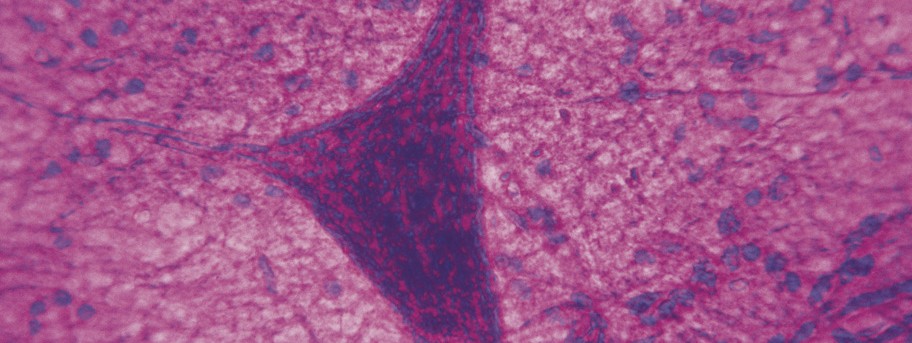
The white matter of the brain is made up of billions of microscopic nerve fibres that pass information in the form of tiny electrical signals. To increase the speed at which these signals travel, each nerve fibre is encased by a sheath formed from a fatty substance, called myelin. Previous studies have shown that the appearance of white matter in magnetic resonance images depends on the angle between the nerve fibres and the direction of the very strong magnetic field used in an MRI scanner.
Based on knowledge of the molecular structure of myelin, the Nottingham physicists devised a new model in which the nerve fibres are represented as long thin hollow tubes with special (anisotropic) magnetic properties. This model explains the dependence of image contrast on fibre orientation in white matter and potentially allows information about the nerve fibres (such as their size and direction) to be inferred from magnetic resonance images.
Research Fellow Dr Samuel Wharton said: “While most MRI-based research focuses on tissue measurements at the millimetre length scale, our experimental scans on healthy human volunteers and modelling of the myelin sheath shows that much more detailed microscopic information relating to the size and direction of nerve fibres can be generated using fairly simple imaging techniques. The results will give clinicians more context in which to recognise and identify lesions or abnormalities in the brain and will also help them to tailor different types of scan to a particular patient.”
Head of the School of Physics and Astronomy, Professor Richard Bowtell added “These results should be an important boost to the world of biomedical imaging which is a key research priority here at The University of Nottingham. We have a strong heritage of groundbreaking work in MRI at the Sir Peter Mansfield Magnetic Resonance Centre and the work was carried out using our 7T scanner which is the strongest magnetic field system for scanning human subjects in the UK.”
Dr Nikolaos Evangelou, Clinical Associate Professor specialising in multiple sclerosis at the Nottingham University Hospitals Trust said: “This research opens new avenues of looking at the nerve fibres in the brain. The more we understand about the nerves and the myelin around them, the more successful we are in studying brain diseases, such as multiple sclerosis. The recent advances in our understanding and treatments of MS are based on basic, solid research such as the one presented by Dr Wharton and Prof Bowtell.
The research will give scientists and clinicians all over the world a better understanding of the effects of nerve fibres and their orientation in magnetic resonance imaging and has potentially useful applications in the diagnosis and monitoring of brain and nervous system diseases like multiple sclerosis where there are known links to myelin loss.
The full research paper ‘Fiber orientation-dependent white matter contrast in gradient echo MRI’ by Dr Samuel Wharton and Professor Richard Bowtell is available here.
Tags: brain, MRI, myelin, PNAS, School of Physics and Astronomy, Sir Peter Mansfield Magnetic Resonance Centre
Leave a Reply
Other News

Top prize for quantum physicist
A University of Nottingham physicist has won a prestigious medal from the Institute of Physics for […]

Zero carbon HOUSE designed and built by students comes home
Design and construct a low cost, zero carbon, family starter home, transport it to Spain, build […]